5. Axial muscles of the abdominal wall and thorax
It is a complex job to balance the body on two feet and walk upright. The muscles of the vertebral column, thorax, and abdominal wall extend, flex, and stabilize different parts of the body’s trunk. The deep muscles of the body’s core help maintain posture as well as provide stability for movement of the limbs.
Muscles of the Abdomen
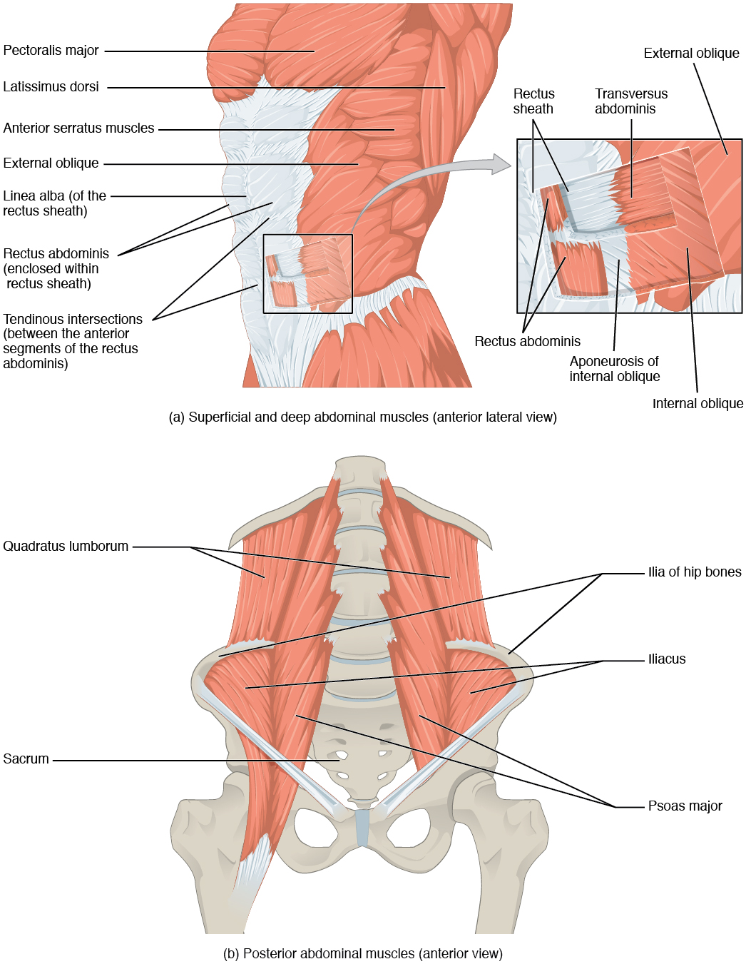
There are four pairs of abdominal muscles that make up the abdominal wall: the rectus abdominis, the external abdominal obliques, the internal abdominal obliques and the transverse abdominis (Figure 5.1 and Table 11.6).
| Muscles of the Abdomen (Table 11.6) | |||||
|---|---|---|---|---|---|
| Movement | Target | Target motion direction | Prime mover | Origin | Insertion |
| Twisting at waist; also bending to the side | Vertebral column | Supination; lateral flexion | External obliques; internal obliques | Ribs 5–12; ilium | Ribs 7–10; linea alba; ilium |
| Squeezing abdomen during forceful exhalations, defecation, urination, and childbirth | Abdominal cavity | Compression | Transversus abdominus | Ilium; ribs 5–10 | Sternum; linea alba; pubis |
| Sitting up | Vertebral column | Flexion | Rectus abdominis | Pubis | Sternum; ribs 5 and 7 |
| Bending to the side | Vertebral column | Lateral flexion | Quadratus lumborum | Ilium; ribs 5–10 | Rib 12; vertebrae L1–L4 |
Muscles of the Abdomen
| Prime mover | Movement | Target | Target motion direction | Origin | Insertion |
|---|---|---|---|---|---|
| External obliques; internal obliques | Twisting at waist; also bending to the side | Vertebral column | Supination; lateral flexion | Ribs 5–12; ilium | Ribs 7–10; linea alba; ilium |
| Transversus abdominus | Squeezing abdomen during forceful exhalations, defecation, urination, and childbirth | Abdominal cavity | Compression | Ilium; ribs 5–10 | Sternum; linea alba; pubis |
| Rectus abdominis | Sitting up | Vertebral column | Flexion | Pubis | Sternum; ribs 5 and 7 |
| Quadratus lumborum | Bending to the side | Vertebral column | Lateral flexion | Ilium; ribs 5–10 | Rib 12; vertebrae L1–L4 |
There are three flat skeletal muscles in the antero-lateral wall of the abdomen. The external oblique, closest to the surface, extend inferiorly and medially, in the direction of sliding one’s four fingers into pants pockets. Perpendicular to it is the intermediate internal oblique, extending superiorly and medially, the direction the thumbs usually go when the other fingers are in the pants pocket. The deep muscle, the transverse abdominis, is arranged transversely around the abdomen, similar to a belt. This arrangement of three bands of muscles in different orientations allows various movements and rotations of the trunk. The three layers of muscle also help to protect the internal abdominal organs in an area where there is no bone.
The linea alba is a white, fibrous band that is made of the bilateral rectus sheaths (see Figure 5.1) that join at the anterior midline of the body. These enclose the rectus abdominis muscles that originate at the pubic crest and symphysis, and extend the length of the body’s trunk. Each muscle is segmented by three transverse bands of collagen fibers called the tendinous intersections resulting in the look of “six-pack abs”.
The posterior abdominal wall is formed by the lumbar vertebrae, parts of the ilia of the hip bones, psoas major and iliacus muscles, and quadratus lumborum muscle. This part of the core plays a key role in stabilizing the rest of the body and maintaining posture.
Muscles of the Thorax
The muscles of the chest serve to facilitate breathing by changing the volume of the thoracic cavity (Table 11.7). When you inhale your chest rises increasing the volume of the thoracic cavity. Alternately, when you exhale, your chest falls decreasing the volume of the thoracic cavity.
| Muscles of the Thorax (Table 11.7) | |||||
|---|---|---|---|---|---|
| Movement | Target | Target motion direction | Prime mover | Origin | Insertion |
| Inhalation; exhalation | Thoracic cavity | Compression; expansion | Diaphragm | Sternum; ribs 6–12; lumbar vertebrae | Central tendon |
| Inhalation;exhalation | Ribs | Elevation (expands thoracic cavity) | External intercostals | Rib superior to each intercostal muscle | Rib inferior to each intercostal muscle |
| Forced exhalation | Ribs | Movement along superior/inferior axis to bring ribs closer together | Internal intercostals | Rib inferior to each intercostal muscle | Rib superior to each intercostal muscle |
Muscles of the Thorax
| Prime mover | Movement | Target | Target motion direction | Origin | Insertion |
|---|---|---|---|---|---|
| Diaphragm | Inhalation; exhalation | Thoracic cavity | Compression; expansion | Sternum; ribs 6–12; lumbar vertebrae | Central tendon |
| External intercostals | Inhalation;exhalation | Ribs | Elevation (expands thoracic cavity) | Rib superior to each intercostal muscle | Rib inferior to each intercostal muscle |
| Internal intercostals | Forced exhalation | Ribs | Movement along superior/inferior axis to bring ribs closer together | Rib inferior to each intercostal muscle | Rib superior to each intercostal muscle |
The Diaphragm
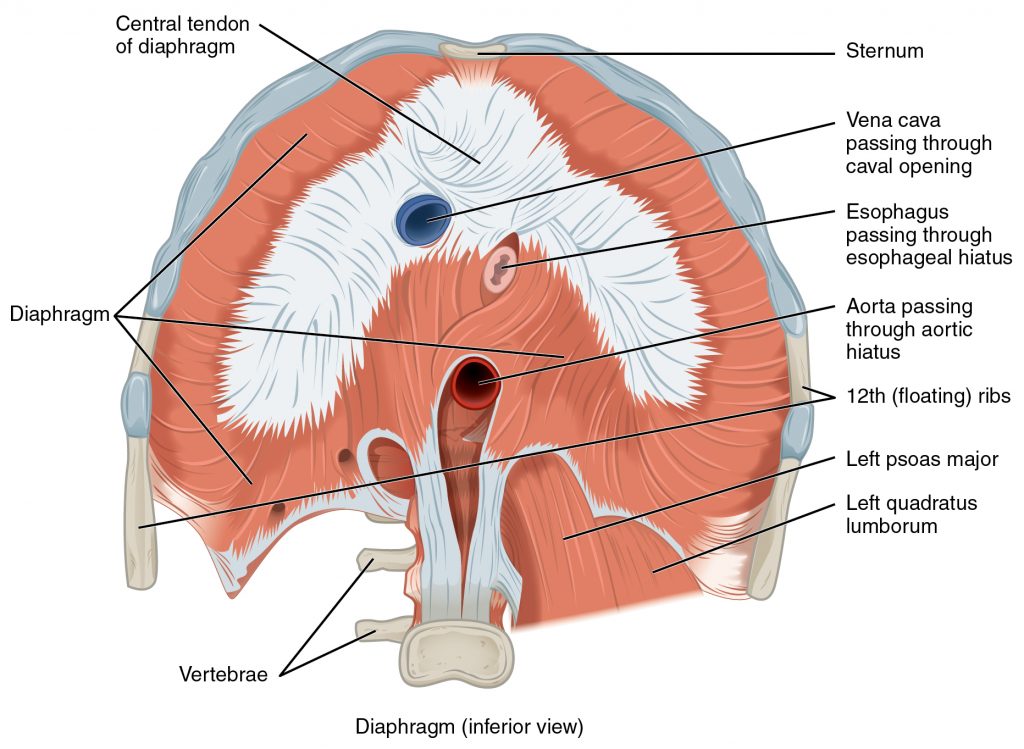
The change in volume of the thoracic cavity during breathing is due to the alternate contraction and relaxation of the diaphragm (Figure 5.2). It separates the thoracic and abdominal cavities, and is dome-shaped at rest. The superior surface of the diaphragm is convex, creating the elevated floor of the thoracic cavity. The inferior surface is concave, creating the curved roof of the abdominal cavity.
Defecating, urination, and even childbirth involve cooperation between the diaphragm and abdominal muscles (this cooperation is referred to as the “Valsalva maneuver”). While you hold your breath the diaphragm and abdominal muscles contract increasing the pressure of the peritoneal cavity and stabilizing the core. When the abdominal muscles contract, the pressure cannot push the diaphragm up, so it increases pressure on the intestinal tract (defecation), urinary tract (urination), or reproductive tract (childbirth).
The inferior surface of the pericardial sac and the inferior surfaces of the pleural membranes (parietal pleura) fuse onto the central tendon of the diaphragm. To the sides of the tendon are the skeletal muscle portions of the diaphragm, which insert into the tendon while having a number of origins including the xiphoid process of the sternum anteriorly, the inferior six ribs and their cartilages laterally, and the lumbar vertebrae and 12th ribs posteriorly.
The diaphragm also includes three openings for the passage of structures between the thorax and the abdomen. The inferior vena cava passes through the caval opening, and the esophagus and attached nerves pass through the esophageal hiatus. The aorta, thoracic duct, and azygous vein pass through the aortic hiatus of the posterior diaphragm.
The Intercostal Muscles
There are three sets of muscles, called intercostal muscles, which span each of the intercostal spaces. The principal role of the intercostal muscles is to assist in breathing by changing the dimensions of the rib cage (Figure 5.3).
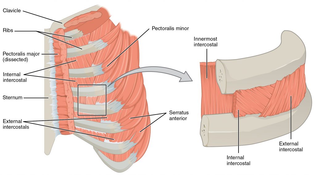
The 11 pairs of superficial external intercostal muscles aid in inspiration of air during breathing because when they contract, they raise the rib cage, which expands it. The 11 pairs of internal intercostal muscles, just under the externals, are used for expiration because they draw the ribs together to constrict the rib cage. The innermost intercostal muscles are the deepest, and they act as synergists for the action of the internal intercostals.
Muscles of the Pelvic Floor and Perineum
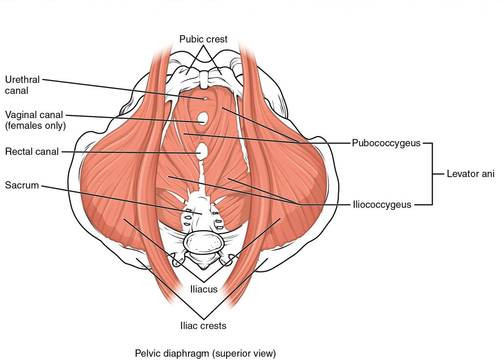
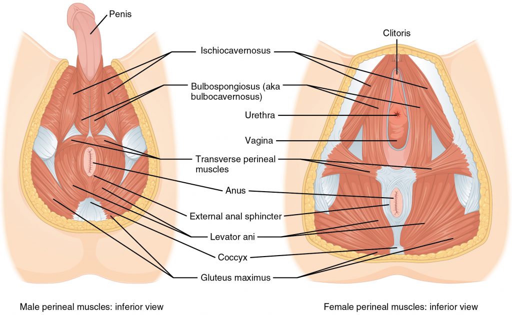
The pelvic floor (also referred to as the pelvic diaphragm) is a muscular sheet that defines the inferior portion of the pelvic cavity. The pelvic floor extends anteriorly to posteriorly from the pubis to the coccyx and is comprised of the levator ani and the ischiococcygeus. Its openings include the anal canal and urethra, and the vagina in women.
The large levator ani consists of two skeletal muscles, the pubococcygeus and the iliococcygeus (Figure 5.4). The levator ani is considered the most important muscle of the pelvic floor because it supports the pelvic viscera. It resists the pressure produced by contraction of the abdominal muscles so that the pressure is applied to the colon to aid in defecation and to the uterus to aid in childbirth (assisted by the ischiococcygeus, which pulls the coccyx anteriorly). This muscle also creates skeletal muscle sphincters at the urethra and anus.
The perineum is the diamond-shaped space between the pubic symphysis (anteriorly), the coccyx (posteriorly), and the ischial tuberosities (laterally), lying just inferior to the pelvic diaphragm (levator ani and ischiococcygeus). Divided transversely into triangles, the anterior is the urogenital triangle, which includes the external genitals and the posterior is the anal triangle containing the anus (Figure 5.4). The perineum is also divided into superficial and deep layers with some of the muscles common to men and women (Figure 5.5). Women also have the compressor urethrae and the sphincter urethrovaginalis, which function to close the vagina. In men, the deep transverse perineal muscle plays a role in ejaculation.
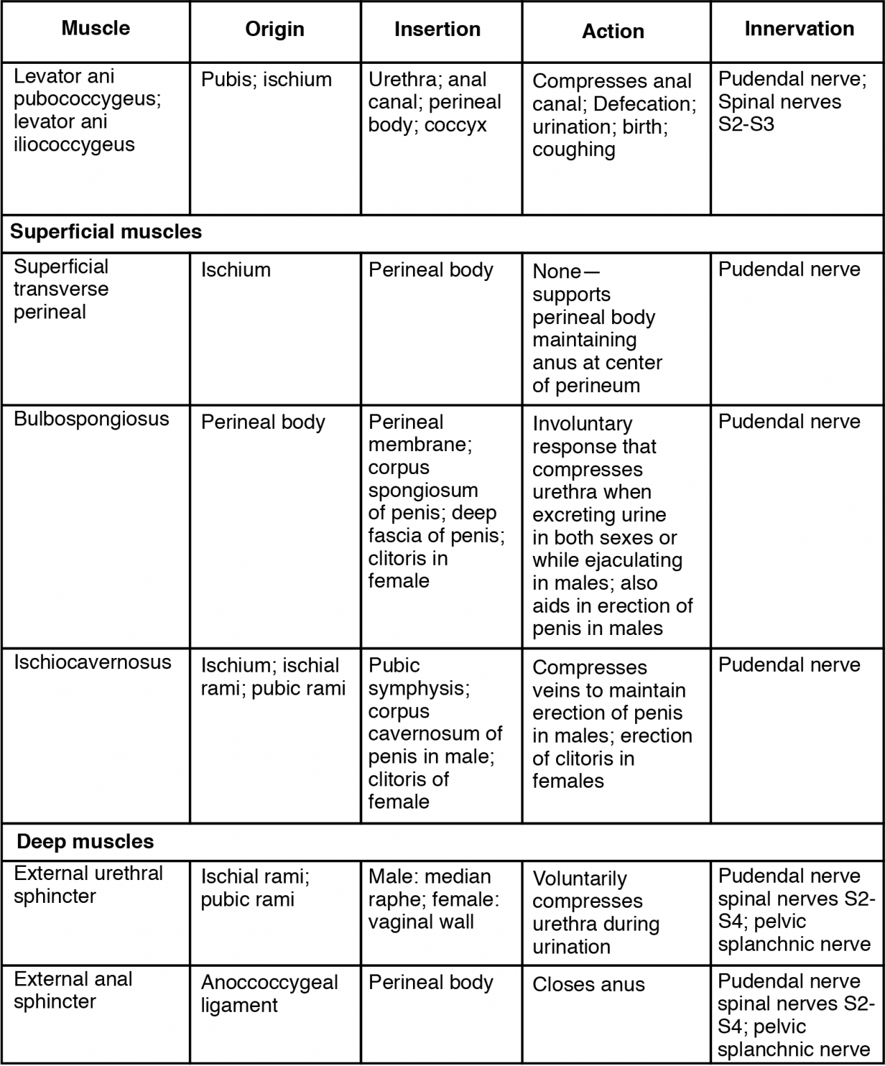
Muscles of the Perineum Common to Men and Women
| Muscle | Origin | Insertion | Action | Innervation |
|---|---|---|---|---|
| Levator ani pubococcygeus; levator ani iliococcygeus | Pubis; ischium | Urethra; anal canal; perineal body; coccyx | Compresses anal canal; defecation; urination; birth; coughing | Pudendal nerve; Spinal nerves S2-S3 |
| Superficial muscles | ||||
| Superficial transverse perineal | Ischium | Perineal body | None- supports perineal body maintaining anus at center of perineum | Pudendal nerve |
| Bulbospongiosus | Perineal body | Perineal membrane; corpus spongiosum of penis; deep fascia of penis; clitoris in female | Involuntary response that compresses urethra when excreting urine in both sexes or while ejaculating in males; also aids in erection of penis in malse | Pudendal nerve |
| Ischiocavernosus | Ischium; ischial rami; pubic rami | Pubic symphysis; corpus cavernosum of penis in males; clitoris in females | Compresses veins to maintain erection of penis in males; erection of clitoris in females | Pudendal nerve |
| Deep muscles | ||||
| External urethral sphincter | Ischial rami; pubic rami | Male: median raphe; female: vaginal wall | Voluntarily compresses urethra during urination | Pudendal nerve spinal nerves S2-S4; pelvic splanchnic nerve |
| External anal sphincter | Anoccoccygeal ligament | Pernieal body | Closes anus | Pudendal nerve spinal nerves S2-S4; pelvic splanchnic nerve |